Skip to content
NEI Data Commons logo
adaptive optics logo
Explore open-source software tools developed for adaptive optics retinal imaging data.

Overview

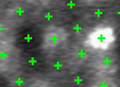
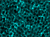
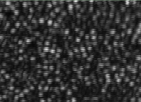
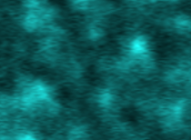
The NEI Clinical and Translational Imaging Section provides custom software for handling, quantifying, and visualizing adaptive optics retinal imaging datasets.

Resources

Image
cone detection logo

Cone Detection blue arrow logo

A software package for identifying cone photoreceptors in non-confocal adaptive optics images such as split detection.
Image
Cone detection ML image

Cone Detection ML blue arrow logo

A software package for detecting cone photoreceptor cells from non-confocal split detection adaptive optics images using machine learning.
Image
cone segmentation logo

Cone Segmentation blue arrow logo

A software package for segmenting the boundaries of cone photoreceptors in non-confocal adaptive optics images such as split detection.
Image
cone segmentation ML image

Cone Segmentation ML blue arrow logo

A software package for segmenting cone photoreceptor cells from non-confocal split detection adaptive optics images using machine learning.
Image
RPE Detection image

RPE Detection blue arrow logo

RPE Detection is a software package for identifying retinal pigment epithelial (RPE) cells in adaptive optics images-enhanced indocyanine green (AO-ICG) images using machine learning.
Image
P-GAN illustration image

P-GAN blue arrow logo

A generative AI model for enhancing the visualization of RPE cells from a single adaptive optics optical coherence tomography acquisition.
Image
RRTGAN image logo

RRTGAN blue arrow logo

A generative artificial intelligence (AI) framework for enhancing the visualization of cone photoreceptor cells from sparsely sampled adaptive optics optical coherence tomography (AOOCT) images.
Image
stratified cycleGAN image

Stratified CycleGAN blue arrow logo

A generative artificial intelligence (AI) model designed to enhance fluorescently labeled retinal pigment epithelial (RPE) cells obtained from late phase indocyanine green (ICG) imaging, transforming them into high-resolution, AI-ICG images.

Questions?

Please reach out to Johnny Tam at johnny@nih.gov if you have questions about these resources.


Explore other Research Areas

patient-reported outcomes with lasik logo
Learn about a clinical trial aimed to validate a survey tool that assesses visual symptoms both before and after LASIK surgery and to identify changes over a short period of time.
PLAE logo
Look up gene expression by retina cell type across different studies, four organisms, and multiple developmental stages.